
During an early pregnancy scan, you might hear the sonographer tell you that there is a corpus luteum in your ovary. Your mind may flicker back to the days when you studied science at school, but the biology of the menstrual cycle is often long forgotten.
Corpus luteum is a latin term, and literally translates as Yellow Body. It is a temporary endocrine structure in the ovary which is involved in ovulation and early pregnancy, and is a normal finding during an ultrasound scan.
During your reproductive years, your body will regularly prepare for pregnancy, whether you’re planning to become pregnant or not. The result of this preparation is a woman’s menstrual cycle.
The menstrual cycle has two phases, the follicular phase and the postovulatory, or luteal, phase. The luteal phase lasts for approximately two weeks. During this time, a corpus luteum forms in the ovary.
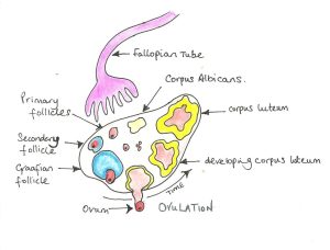
During the follicular phase , the primary follicle forms the secondary follicle and subsequently the mature Graffian or vesicular follicle over approximately 14 days, depending on the length of your menstrual cycle.
At ovulation the follicle ruptures expelling the ovum into the fallopian tube. The remnants of the follicle after ovulation is referred to as the corpus luteum and ranges from 2-5 cm in diameter but involutes as it matures. The corpus luteum produces oestrogen and progesterone, maintaining optimum conditions for implantation if the ovum is fertilised.
The primary purpose of the corpus luteum is to produce hormones, including progesterone. This is known as the luteal phase of the menstrual cycle.
Progesterone is required for a viable pregnancy to occur and to progress. Progesterone enables the endometrium, the uterine lining, to thicken and become spongy. These changes in the uterus allow for implantation of a fertilised egg.
The uterus also provides a rapidly growing embryo with nourishment during its earliest stages of development until the placenta, which also produces progesterone, can take over during the 12th week of pregnancy.
In some instances (especially in twin pregnancies), there may be more than one corpus luteum – (e.g. two corpora lutea)
Many women are unaware of the presence of the corpus luteum, but some may experience mild discomfort on one side. This mild pain will resolve, but can be alleviated by taking paracetamol. Some women may be referred by their doctor for a scan to make sure that the pain is not caused by an ectopic pregnancy.
On an ultrasound scan, the corpus luteum has a typical appearance of a cyst with thick walls, a crenelated inner margin and internal echoes, and a characteristic “ring of fire” appearance of peripheral vascularity when colour Doppler is applied. It usually measures 2 – 3 cm in diameter, but can be bigger. An experienced practitioner can differentiate a corpus luteum from other types of ovarian cyst, giving you the reassurance that this is a normal finding. The corpus luteum reaches a maximum size at about 10 weeks and finally resolves at around 16-20 weeks.
If fertilisation does not occur, or a fertilised egg doesn’t implant in the endometrium, the corpus luteum shrinks away, and progesterone levels drop. The uterine lining is then shed as part of menstruation – the period. The corpus luteum involutes and within a further 14 days will gradually degenerate into a corpus albicans (white body).